Anatomy of Femur Bone
The femur is also called the thigh bone and is the longest and strongest bone of the body. It is composed of an upper end, a lower end and a shaft.
The upper and bears a rounded head, whereas the lower end is widely expanded to from two large condyles. The head is directed medially. The cylindrical shaft is convex forwards.

Upper end of Femur
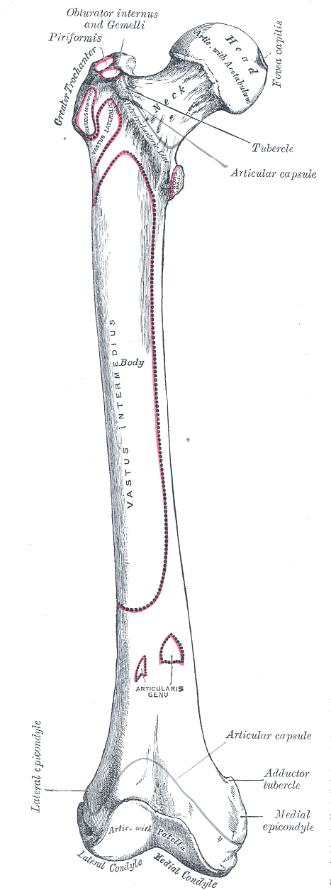
The upper end of the femur includes the head, the neck, the greater trochanter, the lesser trochanter, the intertrochanteric line, and the intertrochanteric crest.
Head of Femur
Head articulates with acetabulum to form a hip joint. It is more than half a sphere and is directed medially, upwards and slightly forwards.
The fovea is a roughened pit just below and behind the center of the head. Head, in its most part, is covered by cartilage.
Neck of Femur
The neck is about is about 3-3.5 cms long and connects head with the shaft. The neck forms an angle with the shaft, known as neck-shaft angle and is about 125 in adults [lesser in females]. The angle facilitates movements of the hip joint. The femoral neck is strengthened by a thickening of bone called the calcar femorale present along its concavity.
The neck has two borders and two surfaces
The upper border, concave and horizontal, meets the shaft at the greater trochanter. The lower border, straight and oblique, meets the shaft near the lesser trochanter.
The anterior surface is flat and meets the shaft at the intertrochanteric line. The anterior surface of the femoral neck is entirely intracapsular. The upper part of this surface may be covered by articular cartilage.
The posterior surface is convex from above downwards and concave from side to side. It meets the shaft at the intertrochanteric crest. It is not intracapsular in its lower lateral part.
Anteversion is the angle formed between the transverse axis of the upper and lower ends of the femur. It is about 15 degrees.
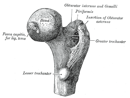
Greater Trochanter
The greater trochanter is a large quadrangular prominence located at the upper part of the junction of the neck with the shaft. The upper border of the trochanter lies at the level of the center of the head.
The greater trochanter has an upper border with an apex, and 3 surfaces (anterior, medial and lateral). The apex is the inturned posterior part of the posterior border. The anterior surface is rough in its lateral part. The medial surface presents a rough impression, above and a deep trochanteric fossa, below. The lateralsurface is crossed by an oblique ridge directed downwards and forwards.
Lesser Trochanter
It is a conical eminence directed medially and backwards from the junction of the posterior part of the neck with the shaft.
Intertrochanteric Line
It marks the junction of the neck with the femur. It is a roughened ridge from the anterosuperior angle of the greater trochanter (as a tubercle) and is continuous below with the spiral line in front of the lesser trochanter.
The spiral line is a curved line with its superior endadjacent to the lesser trochanter, nearly continuouswith the intertrochanteric line, and converging inferiorlywith the pectineal line to form the medial lip of the lineaaspera.
It forms the medial boundary of the distal attachmentof the iliacus muscle. The spiral line winds around the shaft below the lesser trochanter to reach the posterior surface of the shaft.
Intertrochanteric Crest
It marks the junction of the posterior surface of the neck with the shaft of the femur. It is a smooth rounded ridge which begins above at the posterior superiorangle of the greater trochanter and ends at the lesser trochanter. The rounded elevation, a little above its middle is called the quadrate tubercle.
Shaft of Femur
The shaft is almost a cylindrical structure wide superiorly and inferiorly and narrowest in the middle. It is convex forwards and is directed obliquely downwards and medially.

The shaft in middle one-third has three borders -medial, lateral and posterior. The medial and lateral borders are rounded and ill-defined, but the posterior border is in the form of a broad roughened ridge, called the linea aspera. Linea aspera is an important landmark in orthopedics surgeries involving reduction of femoral fractures.
The Linea aspera has distinct medial and lateral lips. The medial and lateral surfaces are directed more backwards than to sides.
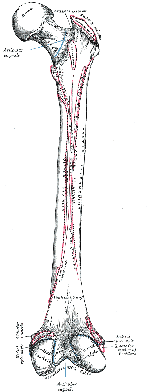
In upper one-third of the shaft, he two lips of the Linea aspera diverge wide to form an additional posterior surface and four borders (medial, lateral, spiral line and the lateral hip of the gluteal tuberosity) and 4 surfaces (anterior, medial, lateral and posterior).
The gluteal tuberosity is a broad roughened ridge on the lateral part of the posterior surface.
Similarly, the two lips of the Linea aspera diverge in lower one third and enclose an additional, popliteal surface. Thus this part of the shaft has four borders (medial, lateral, supracondylar line and lateralsupracondylar line) 4 surfaces (anterior, medial, lateraland popliteal). The medial border and medialsupracondylar line meet inferiorly to obliterate the medial surface.
Lower End of Femur

The lower end of the femur is wide and expanded. It has two large condyles – medial and lateral. Anteriorly, the two condyles are united and are in a line with the front of the shaft. Posteriorly, they are separated by a deep gap, termed the intercondylar fossa or intercondylar notch, and project backwards much beyond the plane of the popliteal surface.
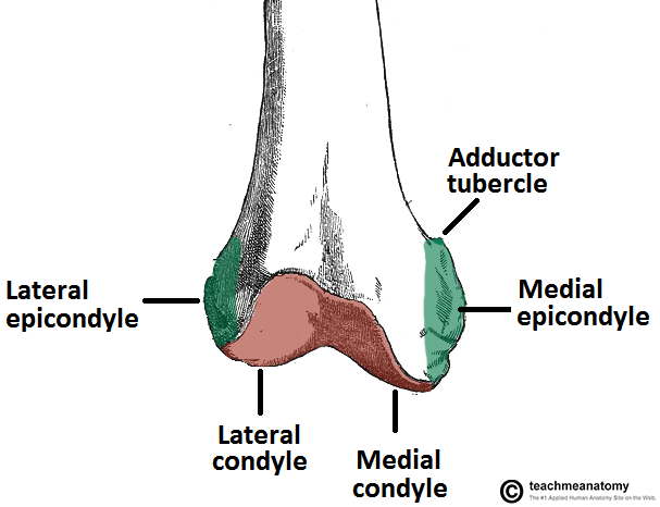

The lateral condyle is flat laterally, less prominent than medial condyle and stouter than it. It has a prominence called the lateral epicondyle. Below it lies the popliteal groove with a deeper anterior part and a shallower posterior part.
Medial condyle is convex medially. It also bears a prominent point called the medial epicondyle. Adductor tubercle is a projection posterosuperior to the epicondyle which serves as an important landmark. The epiphyseal line for the lower end of the femur passes through it.
Intercondylar fossa or notch separates the lower and posterior parts of the two condyles. The intercondylar line separates the notch from the popliteal surface. Anteriorly, the notch is limited by the patellar articular surface.
The two condyles are partially covered by a large articular surface. Anteriorly, the condyles articulate with patella and this articulation extends more on the lateralcondyle than on the medial. Between the two condyles, the surface is grooved vertically. Two faint grooves separate the patellar articulation surface from tibial surfaces. Tibial articulation surface over the lateralcondyle is short and straight anteroposteriorly whereas the part over the medial condyle is longer and is convex medially.
Attachments on the Femur
Head of Femur
The fovea on the head of the femur provides attachment to the ligament of the head (round ligament, or ligamentum teres).
Greater Trochanter
- The piriformis is inserted into the apex
- The gluteus minimus is inserted into the rough lateral part of the anterior surface
- The obturator internus and the two gemelli are inserted into the upper rough impression on the medial surface
- The obturator externus is inserted into the trochanteric fossa
- The gluteus medius is inserted into the ridge on the lateral surface.
- The trochanteric bursa of the gluteus medius lies in front of the ridge, and the trochanteric bursa of the gluteus maximus lies behind the ridge.
Lesser Trochanter
- The psoas major is inserted on the apex and medial part of the rough anterior surface.
- The iliacus is inserted on the anterior surface of the base of the trochanter, and on the area below.
- Gluteus minimus bursa lies deep to the upper horizontal fibers of the adductor magnus.
Intertrochanteric Line
Following structures attach to the intertrochanteric line
- Capsular ligament of the hip joint
- Iliofemoral ligament in its upper part
- Lower band of the iliofemoral ligament in its lower part
- Highest fibers of the vastus lateralis from the upper end
- Highest fibers of the vastus medialis from the lower end
- Quadratus femoris attached on quadrate tubercle
Shaft of Femur
- The medial and popliteal surfaces are bare [ Except for part of gastrocnemius origin on the popliteal surface]
- Vastus intermedius – upper three-fourths of the anterior and lateral surfaces.
- Articularis genu – just below the vastus intermedius.
- Vastus lateralis – upper part of the intertrochanteric line, anterior and inferior borders of the greater trochanter, the lateral lip of the gluteal tuberosity, and the upper half of the laterallip of the line aspera.
- Vastus medialis – Lower part of the intertrochanteric line, the spiral line, the medial lip of the linea aspera, and the upper one –fourth of the medial supracondylar line.
- Gluteal tuberosity receives insertion of deeper fibers of the lower half of the gluteus maximus
- Adductor longus – Medial lip of the linea aspera between the vastus medialis and the adductor brevis and magnus
- Adductor brevis is inserted into a line extending from the lesser trochanter to the upper part of the linea aspera, behind the pectineus and the upper part of the adductor longus.
- Adductor magnus is inserted into the medialmargin of the gluteal tuberosity, the linea aspera, the medial supracondylar line, and the adductor tubercle
- Pectineus is inserted on a line extending from the lesser trochanter to the linea aspera.
- The short head of the biceps femoris arises from the lateral lip of the linea aspera between the vastus lateralis and the adductor magnus, and from the upper two – thirds of the lateralsupracondylar line
- Medial and lateral intermuscular septa are attached to the lips of the linea aspera and to the supracondylar line. These septae separate the extensor muscles from the adductor medially, and from the flexors laterally. The lower end of the lateral supracondylar line gives origin to the plantaris above and the upper part of the lateralhead of the gastrocnemius below.
- The popliteal surface is covered with fat and forms the floor of the popliteal fossa. Medial head of the gastrocnemius extends to the popliteal surface just above the medial condyle.
Lateral Condyle
- Fibular collateral ligament of the knee attaches to the lateral epicondyle.
- The popliteus arises from the deep anterior part of the popliteal groove. When the knee is flexed, the tendon of this muscle lies in the shallow posterior part of the grove.
- The muscular impression near the lateralepicondyle gives origin to the lateral head of the gastrocnemius.
Medial Condyle
- Tibial collateral ligament of the knee – medialepicondyle
- Hamstring part of the adductor magnus – adductor tubercle
Intercondylar Notch
- Anterior cruciate ligament – posterior part of the medial surface of the lateral condyle.
- The intercondylar line provides attachment to the capsular ligament and laterally to the oblique popliteal ligament.
Ossification of Femur
One primary and four secondary centers.

The primary center for the shaft appears in the 7 weeks of intrauterine life.
The secondary centers appear as follows
- Lower end of Femur – At end of the 9th month of intrauterine life
- Head – first six months of life
- Greater trochanter – 4 years
- Lesser trochanter – 12 years
The upper apophyses (lesser trochanter, greater trochanter and head, in that order) fuse with the shaft at about 18 years. The lower epiphysis fuses by the 20th year.
Blood supply of Femur
- The smaller, medial part of the head, near the fovea, is supplied by medial epiphyseal arteries derived from the posterior division of the obturator artery and from the ascending branch of the medial circumflex femoral artery. These arterial twigs enter the acetabular notch and then pass along the round ligament to reach the head.
- The larger, lateral part of the head is supplied by lateral epiphyseal arteries which are derived from the retinacular branches of the medial circumflex femoral artery. This set constitutes the main supply and damage to it results in necrosis of the head of the following fractures of the neck of the femur. After epiphyseal fusion, the lateralepiphyseal arteries anastomose freely with the metaphyseal arteries.
- The intracapsular neck is supplied by the retinacular arteries derived chiefly from the trochanteric anastomosis. The vessels produce longitudinal grooves and foramina directed towards the head, mainly on the anterior and posterior- superior surface. The extracapsular part of the neck is supplied by the ascending branch of the medial circumflex femoral artery.
- Nutrient artery to shaft of the femur is derived from the second perforating artery. The nutrient foramen (or foramina) is located on the medialside on the linea aspera and is directed upwards.
- The lower end is supplied by genicular arteries and anastomosis around the knee
Significance of Femur
- Femur is a common bone to be injured
- Ossification of the lower end of the femur is of medicolegal importance. Presence of its center in a newly born child found dead indicates that the child was capable of independent existence.
- Coxa valga is a condition where the femoral neck-shaft angle is more than normal. ( 135 degrees)
- Coxa vara is a condition where the neck-shaft angle is less than normal (120 degrees)


Comments
Post a Comment