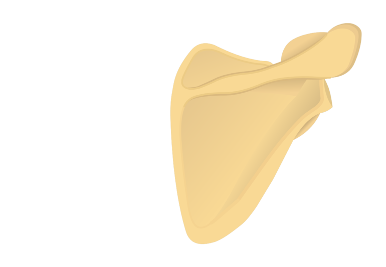
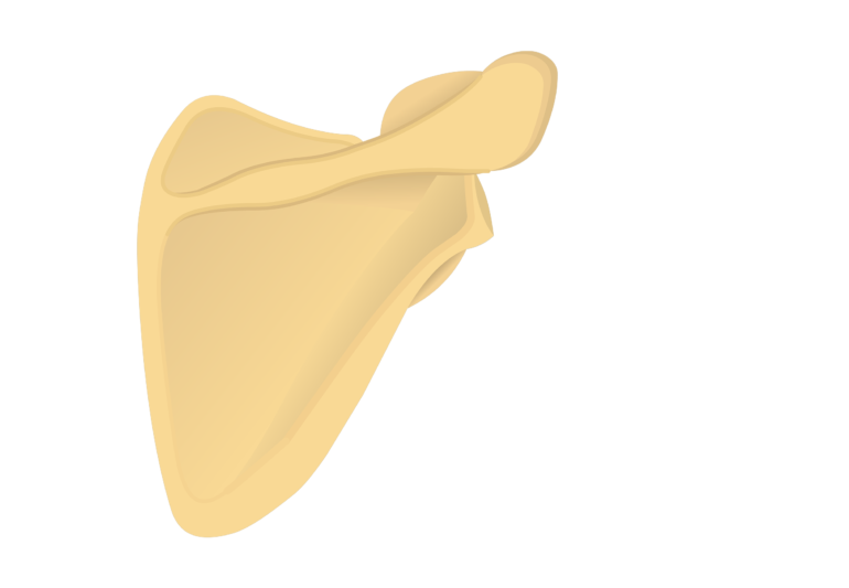
Introduction to Scapula Bone:
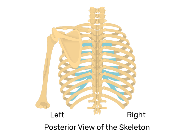
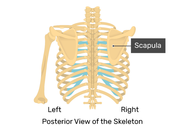
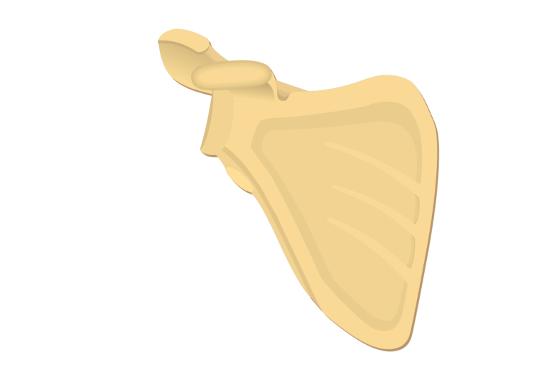
The scapula or shoulder blade is a flat, triangular-shaped bone that to the posterior surface of ribs 2-7.
Markings of the Scapula Bone:
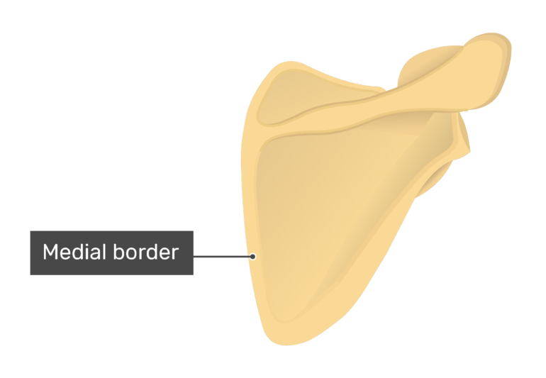
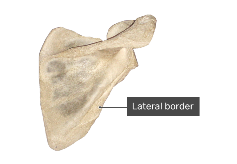
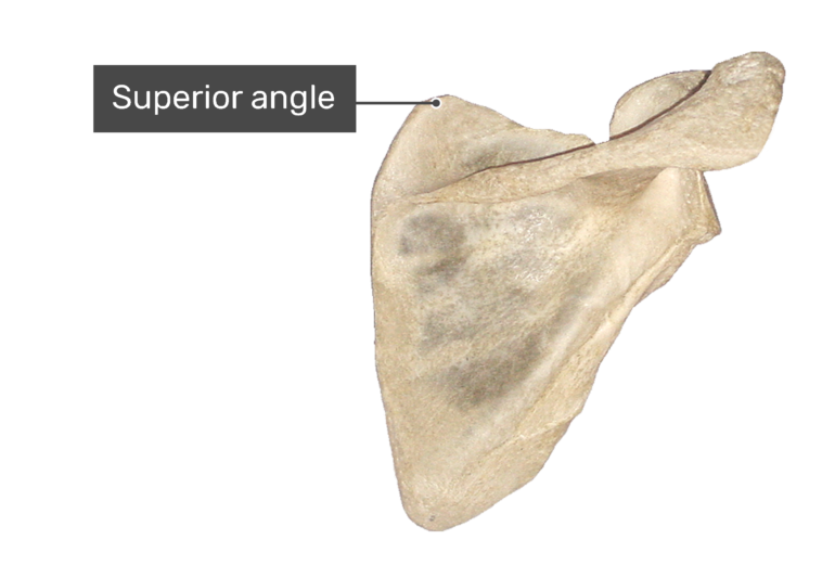
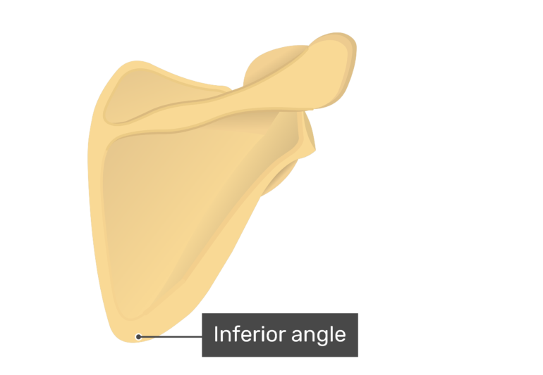
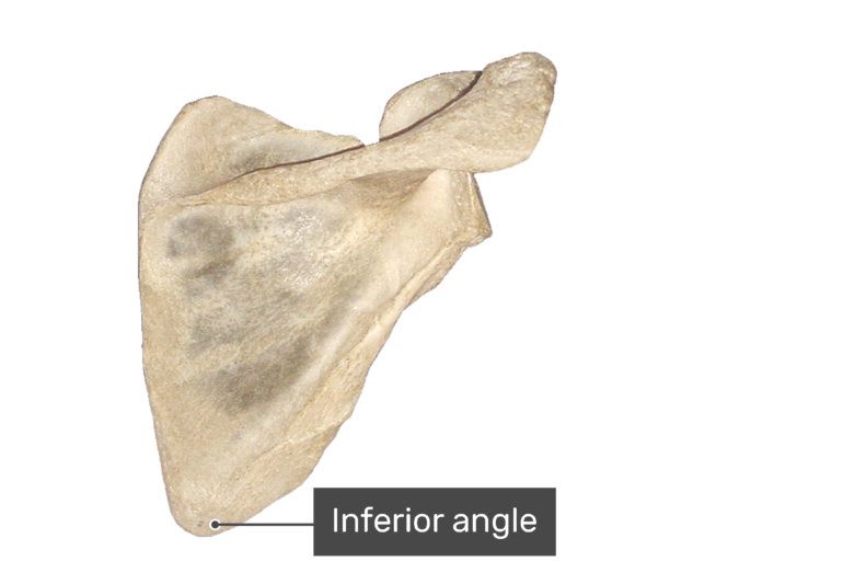
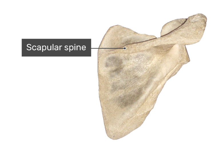
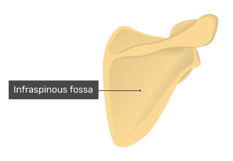
- It has three borders (superior, lateral, medial), three angles (superior, lateral, medial) and two surfaces (costal, dorsal). A prominent ridge or spine divides the dorsal surface into two, unequal parts called the supraspinous fossa and infraspinous fossa.
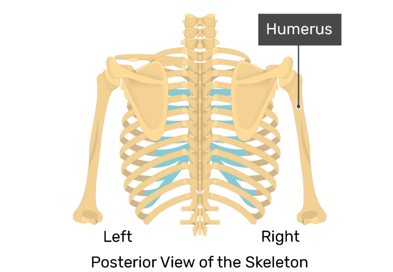
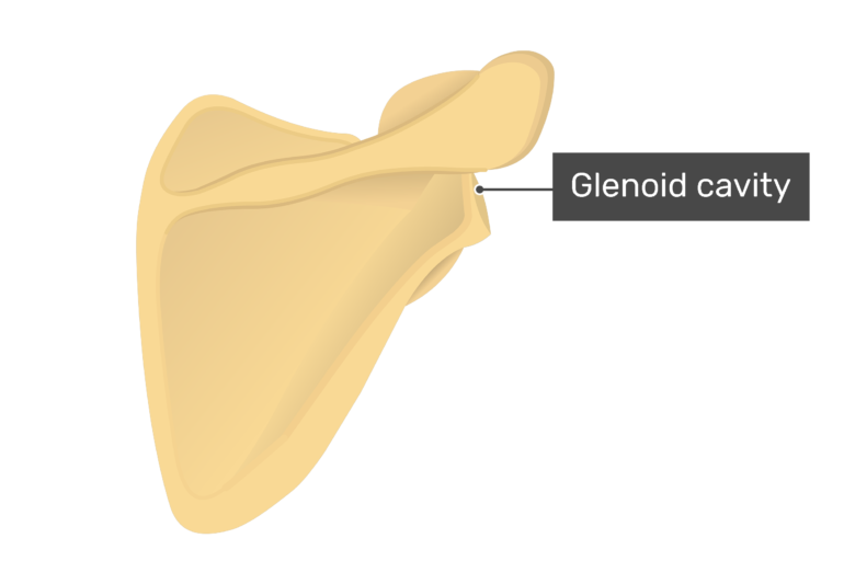
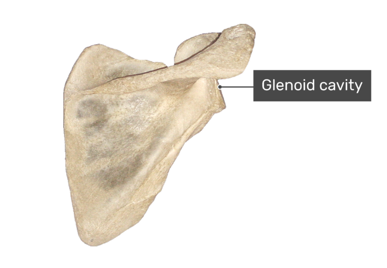
- A shallow depression at the lateral angle called the glenoid cavity the head of the humerus to form the shoulder or gleno-humeral joint.
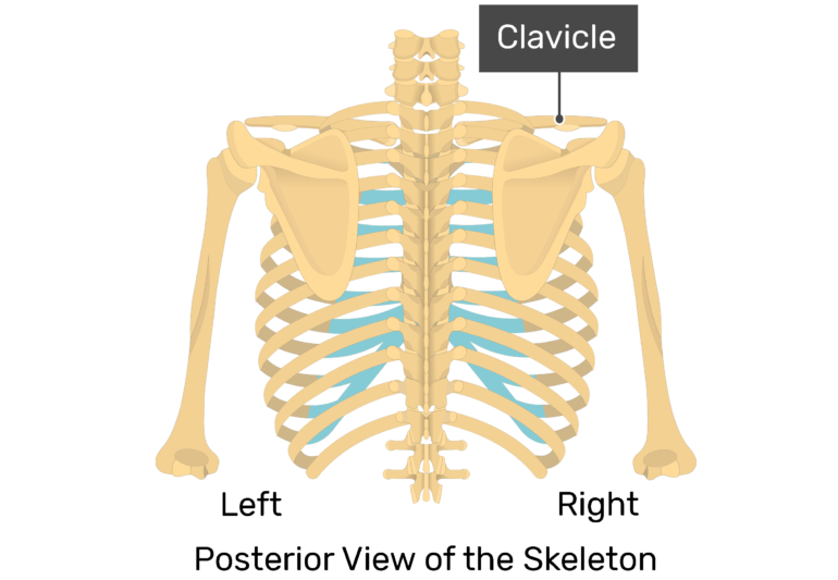
3.The scapula also with the clavicle to form the shoulder girdle or pectoral girdle, which supports movements of the humerus.

4.Seventeen to the borders, angles, ridges, bumps, processes, and fossae found on the surface of the scapula.

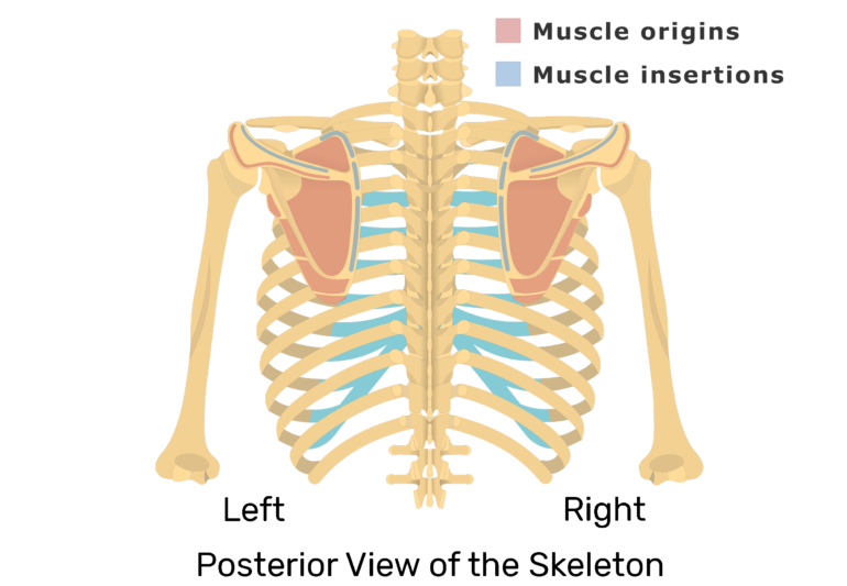
5.The muscles that insert on the scapula (intrinsic muscles) connect it to the axial skeleton. When these muscles contract, the scapula is elevated, depressed, rotated, protracted, retracted, or stabilized when the humerus is moved.
6.The muscles that originate on the scapula (extrinsic muscles) insert on the humerus, radius, and ulna. They cause the humerus to rotate, abduct, adduct, flex or extend.
Anterior Scapula Bone:
Bone Markings:
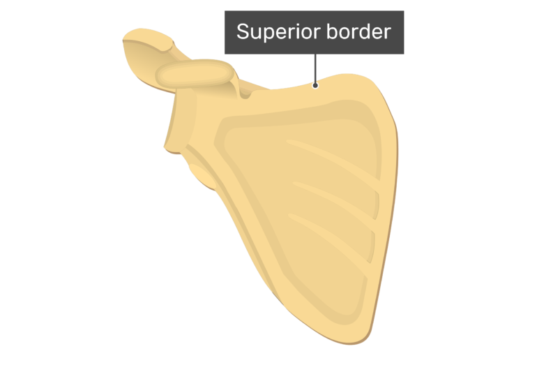
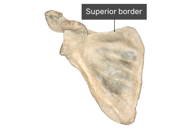
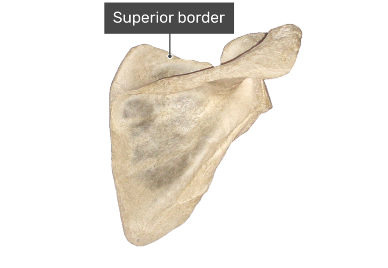
- Superior border or margin (Margo superior) is the upper edge of the scapula that runs next to the clavicle. The omohyoid muscle attaches along this surface.
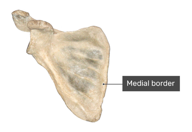
(Margo medialis) is the medial edge of the scapula and is also called the vetebral border. This area is an attachment point for the rhomboid major, rhomboid minor and serratus anterior muscles.


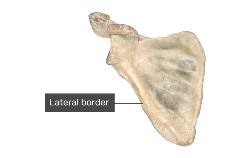
- (Margo lateralis) is the lateral edge of the scapula. The teres minor muscle attaches along this surface, which is also called the axillary border.


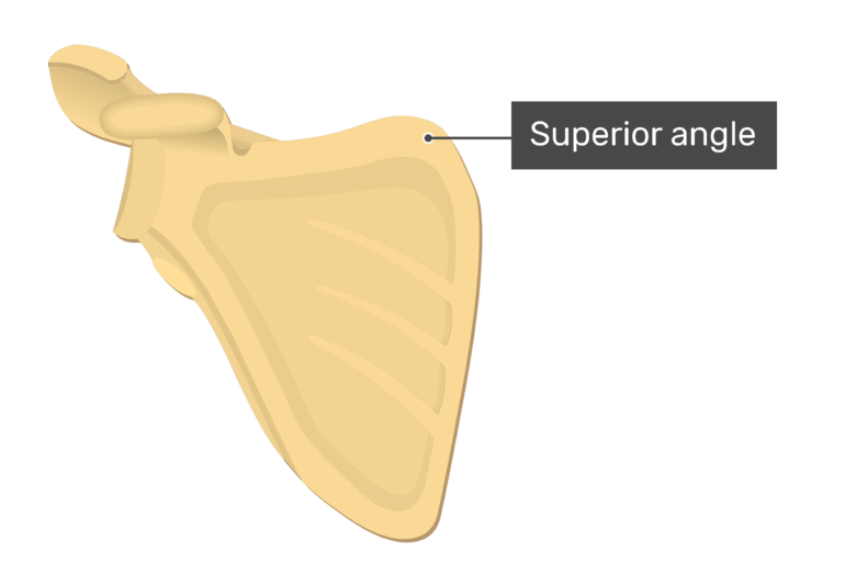
(Angulus superior) is a sharp curvature at the junction of the superior and medial borders. This area is an attachment point for levator scapula muscle.

(Angulus lateralis) is the junction of the superior and lateral borders. Its slightly concave lateral edge articulates with the head of the humerus.


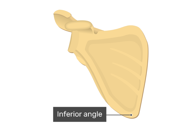
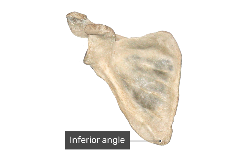
(Angulus inferior) is a sharp curvature formed at the junction of the medial and lateral margins. It is an area of attachment for the teres major muscle.
(Processus coracoi-deus) is a curved, hook-like anterolateral projection located above the lateral angle.
- The term coracoid refers to the marking’s resemblance to a crow’s beak (Gr., korax, crow’s). The biceps brachii, coracobrachialis, and pectoralis minor muscles attach along the surface of this process.


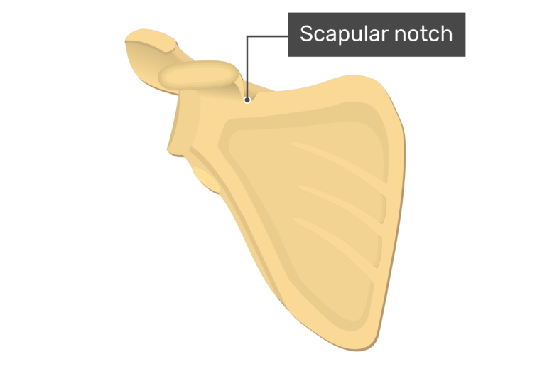
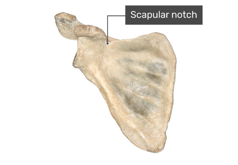
(Incisura scapulae) is a small groove located medial to the coracoid process. It forms a passageway for the suprascapular nerve.
(Cavitas glen-oidalis) is a slight concavity at the lateral angle. It forms a shallow socket for the articulation of the head of the humerus.


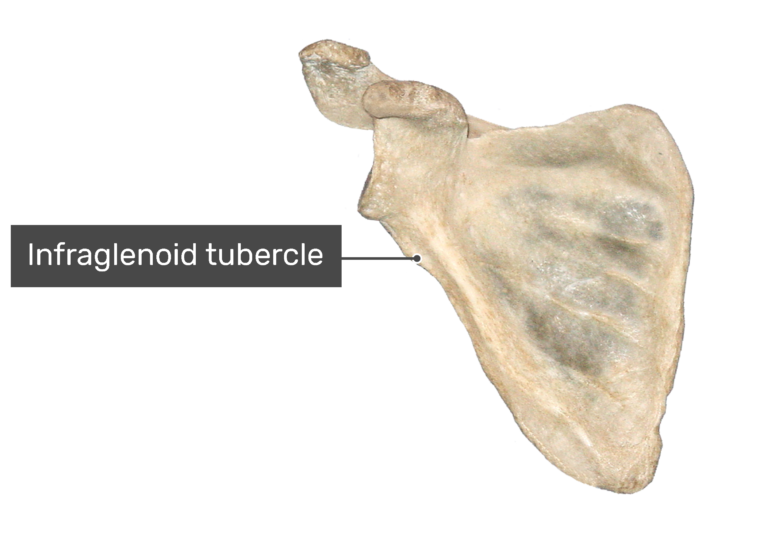
(Tuberculum infra-glenoidale) is a small prominence on the inferior margin of the glenoid fossa. It serves as an attachment point for the long head of the triceps brachii muscle.

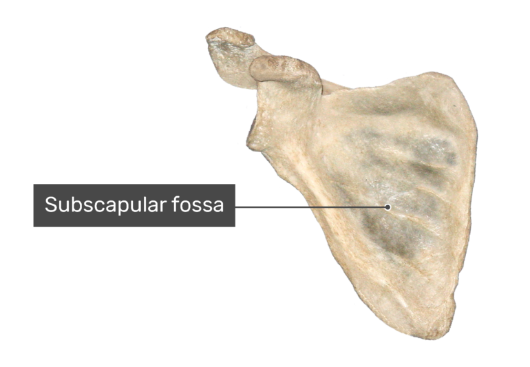
(Fossa subscapularis) is a large, slightly depressed region in the middle of scapula’s anterior surface. The subscapularis muscle attaches to this region of the bone.


Comments
Post a Comment